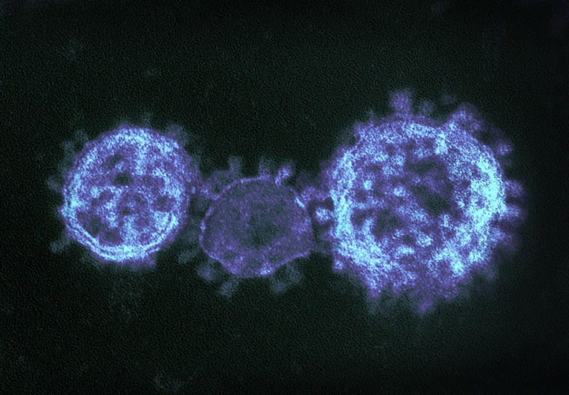
Coronaviruses

Coronaviruses are a large family of viruses that can infect a range of hosts. They are known to cause diseases including the common cold, Severe Acute Respiratory Syndrome (SARS) and Middle East Respiratory Syndrome (MERS) in humans.
In January 2020, China saw an outbreak of a new coronavirus strain now named SARS-CoV-2. Although the animal reservoir for the SARS and MERS viruses are known, this has yet to have been confirmed for SARS-CoV-2. All three strains are transmissible between humans.
To allow the widest possible distribution of relevant research, the Microbiology Society has brought together articles from across our portfolio and made this content freely available.
Image credit: "MERS-CoV" by NIAID is licensed under CC BY 2.0, this image has been modified.
Collection Contents
281 - 298 of 298 results
-
-
Genetic Variation of Neurotropic and Non-neurotropic Murine Coronaviruses
More LessSUMMARYThe murine coronavirus strains MHV JHM, MHV 1, MHV 2, MHV 3 and MHV A59 were tested for their neurovirulence in weanling rats. The strain JHM was found to be highly neurovirulent for weanling rats, whereas the other strains were not, or only slightly, neurovirulent. MHV 1 caused no lesions in weanling rats. The other strains (MHV 2, MHV 3 and MHV A59) induced predominantly subclinical infections in weanling rats as demonstrated by an increase of antibodies and inflammatory lesions in the liver. Analysis of these strains by cross-neutralization revealed variable degrees of antigenic relationship between these viruses which were not related to their neurovirulence. However, when these strains were compared by analysing the T1-RNase-resistant oligonucleotides of virion RNA, the highly neurovirulent strain JHM was found to differ significantly in its nucleotide sequence from the other less-neurovirulent strains.
-
-
-
The Distribution of Human Coronavirus Strain 229E on the Surface of Human Diploid Cells
More LessSUMMARYThe distribution of human coronavirus strain 229E (HCV 229E) particles on the surface of human diploid (MRCc) cells was examined. Virus particles showed a totally random distribution on fixed cells and on cells to which virus had been adsorbed in the cold. A marked redistribution of virus particles was observed on warming virus—cell preparations to 33 °C for 20 min, the peripheral areas of the cell becoming relatively devoid of virus particles while the majority of particles were now located some distance from the edge of the cell. Redistribution did not occur in the presence of metabolic inhibitors.
-
-
-
Coronavirus JHM: Intracellular Protein Synthesis
More LessSummaryCoronavirus JHM contained six major proteins, four of which were glycosylated. The proteins were gp170, gp98, gp65, p60, gp25 and p23. Sac(-) cells infected with JHM shut off host cell protein synthesis, and the synthesis of three major (150K, 60K and 23K) and three minor (65K, 30K and 14K) polypeptides was detected by pulse-labelling with 35S-methionine. Antiserum directed against purified virus proteins specifically immunoprecipitated the three major intracellular species and also the 65K minor species. During a chase period, species 150K and 23K were processed and three new immunoprecipitable species, 170K, 98K and 25K appeared. The intracellular species 170K, 98K, 65K, 60K, 25K and 23K co-electrophoresed with virion proteins.
Two-dimensional gel electrophoresis of infected cell polypeptides showed that the 60K, 23K, 25K and 14K species were relatively basic polypeptides whilst the 98K and 170K were relatively acidic and heterogeneously charged polypeptides. Additionally, a charge-size modification of the 23K species during processing was detected, which was not apparent using one-dimensional gel analysis.
-
-
-
Structural Polypeptides of Coronavirus IBV
More LessSummaryAvian infectious bronchitis virus (IBV) was grown and radiolabelled with 35S-methionine, 3H-leucine and 3H-glucosamine in de-embryonated chicken eggs. Approximately 12 different polypeptides were clearly detected by SDS-polyacrylamide gel electrophoresis of virus preparations. Growth of IBV in chorioallantoic membrane cells labelled with 35S-methionine indicated that most of these polypeptides, and additional ones, some of which were glycosylated, were host components. Five polypeptides appeared to be virus-coded, with apparent mol. wt. of 94 × 103, 84 × 103, 54 × 103, 30 × 103 and 28 × 103. Four of these, p94, p84, p30 and p28, were glycosylated. The virion spikes appeared to be composed of p94 and p84, while p30 and p28 were partially embedded in the virion membrane. By analogy with other reports, p54 is the nucleocapsid polypeptide.
-
-
-
Coronavirus JHM: a Virion-associated Protein Kinase
More LessSUMMARYCoronavirus JHM contains six major proteins, one of which, the 60000 mol. wt. nucleocapsid protein pp60, is phosphorylated. In JHM-infected cells ip 60K, the intracellular precursor to pp60 is also phosphorylated. Associated with purified JHM virions is a protein kinase which will phosphorylate pp60 and a variety of exogenous substrates in vitro. The enzyme has the characteristics of a cyclic nucleotide-independent protein kinase. Both the in vivo reaction and the enzyme activity in vitro transferred the γ-phosphate of ATP to serine residues on the nucleocapsid protein.
-
-
-
The Polypeptide Structure of Canine Coronavirus and its Relationship to Porcine Transmissible Gastroenteritis Virus
More LessSUMMARYCanine coronavirus (CCV) isolate 1–71 was grown in secondary dog kidney cells and purified by rate zonal centrifugation. Polyacrylamide gel electrophoresis revealed four major structural polypeptides with apparent mol. wt. of 203800 (gp204), 49800 (p50), 31800 (gp32) and 21600 (gp22). Incorporation of 3H-glucosamine into gp204, gp32 and gp22 indicated that these were glycopolypeptides. Comparison of the structural polypeptides of CCV and porcine transmissible gastroenteritis virus (TGEV) by co-electrophoresis demonstrated that TGEV polypeptides corresponded closely, but not identically, with gp204, p50 and gp32 of CCV and confirmed that gp22 was a major structural component only in the canine virus. The close similarities in structure of the two coronaviruses augments the relationship established by serology.
-
-
-
Enzyme-linked Immunosorbent Assay for Coronaviruses HCV 229E and MHV 3
More LessSUMMARYThe antigenic relationship between human coronavirus strain 229E (HCV 229E) and mouse hepatitis virus strain 3 (MHV 3) was studied by means of the indirect form of enzyme-linked immunosorbent assay (ELISA). A cross-reaction was found with hyperimmune rabbit sera between HCV 229E and MHV 3 which may be due to the adherence of bovine serum components from tissue culture media, which were present on virus particles even after extensive purification. No cross-reaction was observed with immune sera absorbed with bovine serum, or with HCV 229E grown in tissue culture without serum. This indirect ELISA with HCV 229E may prove to be useful for studies with human sera.
-
-
-
Structural Polypeptides of the Murine Coronavirus JHM
More LessSUMMARYAnalysis by SDS-polyacrylamide gel electrophoresis shows that the purified coronavirus JHM contains six polypeptides. The apparent mol. wt. of the polypeptides (GP1, GP2, GP3, VP4, GP5 and VP6) are 170000; 125000; 97500; 60800; 24800 and 22700, respectively. Four polypeptides are glycosylated (GP1, GP2, GP3 and GP5). The analysis of particles obtained after limited proteolysis with pronase suggests that GP2 and GP3 are protruding from the lipid envelope and, together with GP1, form the spike layer. Protein VP6 and a part of GP5 are located within the lipid bilayer. Protein VP4 is susceptible to digestion at a concentration of pronase which changes the morphology of the virus particles making the interior of the virus accessible. Subviral particles produced after treatment with the detergent Nonidet P40 banded at a higher density than the virus and contained only VP4, GP5 and VP6.
-
-
-
Genomic RNA of the Murine Coronavirus JHM
More LessSUMMARYGenomic RNA extracted from the purified murine coronavirus JHM sediments between 52S and 54S in aqueous sucrose gradients. The RNA is single-stranded and has an apparent mol. wt. of 5·4 to 6·5 ×106, as determined by electrophoresis in polyacrylamide agarose gels of different concentrations. The presence of polyadenylate sequences in the RNA is demonstrated by binding to oligo-(dT) cellulose and digestion with ribonucleases A and T1. The purified RNA does not dissociate into subunits at high temperatures or in high concentrations of DMSO and is infectious.
-
-
-
Ribonucleoprotein-like Structures from Coronavirus Particles
More LessSUMMARYThe structure of the ribonucleoprotein (RNP) complex of three coronaviruses was investigated. A single-stranded helix of diam. 14 to 16 nm and up to 320 nm in length was released from disrupted particles of human coronavirus strain 229E and mouse hepatitis virus strain 3 after incubation in mild conditions. The helical complexes appeared to be composed of globular subunits with long axes of 5 to 7 nm surrounding a hollow core of diam. 3 to 4 nm. The complexes were shown to be sensitive to both pancreatic RNase and to pronase. No undegraded internal component was obtained from disrupted avian infectious bronchitis virus particles. We conclude that these structures are RNP complexes. The similarity between these RNPs and those of other large lipid containing RNA viruses is discussed.
-
-
-
The Genome of Human Coronavirus Strain 229E
More LessSUMMARYThe genomic RNA of human coronavirus strain 229E (HCV 229E) migrated on polyacrylamide gels as a single peak with a mol. wt. of 5.8 × 10⁶. Denaturation of the genome with formaldehyde did not alter its electrophoretic mobility, which suggests that the HCV 229E genome is a single-stranded molecule. At least 30% of the genomic RNA was shown to contain covalently attached polyadenylic acid [poly(A)] sequences by binding the RNA to an oligo(dT)-cellulose column. These poly(A) tracts were shown to be about 70 nucleotides in length by measuring the resistance to digestion of HCV 229E RNA with pancreatic and T1 RNases. Finally, the genomic RNA was shown to terminate at or near the 3′-terminus on the basis of its susceptibility to polynucleotide phosphorylase.
-
-
-
Presence of Genomic Polyadenylate and Absence of Detectable Virion Transcriptase in Human Coronavirus OC-43
More LessSUMMARYHuman coronavirus RNA, prepared by extraction of purified virions with phenol-chloroform, consists of a major 15 to 55S class and a minor 4S class of RNA fragments. Polyadenylic acid [poly (A)] sequences are present in 15 to 55S but not in 4S RNA, suggesting different functions for each class. A stretch of poly (A) of approximately 19 adenosine monophosphate residues was obtained in sizing experiments after digesting OC-43 RNA with pancreatic and T1 ribonucleases. An OC-43 virion RNA transcriptase could not be detected with systems optimal for detecting the transcriptases of influenza and Newcastle disease virus.
-
-
-
Biological Properties of Avian Coronavirus RNA
More LessSUMMARYRNA with a sedimentation coefficient of 64S was isolated from infectious bronchitis virus, an avian coronavirus. The RNA contained a polyadenylic acid tract and was found to be infectious.
-
-
-
Studies on the Structure of a Coronavirus-Avian Infectious Bronchitis Virus
More LessSUMMARYWhen avian infectious bronchitis virus (IBV) is fixed in formaldehyde, negative stain is able to penetrate the particle and an internal component is visualized. This component is seen as a tongue or flask shaped structure attached at one point to the outer virus membrane. A model yielding transmission patterns similar to the virus has been made. Gradient centrifugation studies on IBV reveal that the RNP is associated with the internal sac.
-
-
-
Haemagglutination by Avian Infectious Bronchitis Virus – a Coronavirus
More LessSummaryThe haemagglutinating ability of three strains of IBV was investigated. It was shown that whereas strain Beaudette had no detectable haemagglutinin, both Connecticut and Massachusetts agglutinated red cells of various species. The haemagglutinin of Connecticut was detectable after sucrose gradient purification whereas that of Massachusetts required both the purification step and incubation with the enzyme phospholipase C to reveal it. The agglutination could be inhibited by specific antisera. Some studies on the nature of the red cell receptor, and the possible presence of a receptor destroying enzyme, are reported.
-
-
-
Electron Microscopic Studies of Coronavirus
More LessSUMMARYElectron-microscopic studies of the morphology and development of a coronavirus (linder strain), isolated in human foetal diploid lung cells from a case of upper respiratory illness, revealed virus particles of diameter 80 to 160 nm. They were initially observed 12 hr after infection. Extracellular and intracytoplasmic virus concentration increased sharply at 18 hr and reached a maximum at 24 hr. The number of particles decreased slightly at 48 hr and by 72 hr many cells were lysed. The particles formed by budding into the cisternae of the endoplasmic reticulum or into an inclusion composed of tubular structures resembling part of the complete virus particle. There were cytoplasmic inclusions of dense particles within a granular matrix and surrounded by a double membrane. The release of particles by lysis is illustrated. Extracellular virus was specifically tagged with ferritin-labelled antibody.
-
-
-
The Morphological and Biological Effects of Various Antisera on Avian Infectious Bronchitis Virus
More LessSummaryBiologically, homotypic and heterotypic antisera neutralized avian infectious bronchitis virus significantly more when unheated. Morphologically, using the electron microscope technique of negative staining, there was a clear distinction between the effects of homotypic and heterotypic antisera. Heated homotypic antiserum revealed antibody attached only to the projections of the virus, while with unheated homotypic serum heat labile components could be visualized but no basic change could be seen in particle morphology. Heterotypic serum contained antibodies directed both against the projections and the envelope of the virus. In addition, unheated heterotypic antiserum produced holes approximately 100 Å in diameter in the virus membrane, suggesting that a form of virus lysis takes place. Rabbit antiserum prepared against uninfected chick-embryo fibroblasts was able to produce similar holes in the virus envelope and this led us to postulate that the envelope component of avian infectious bronchitis virus is closely related to normal chick host material.
-
-
-
The Morphology of Three Previously Uncharacterized Human Respiratory Viruses that Grow in Organ Culture
More LessSummaryA simple method is described for examining organ cultures by electron microscopy for the presence of virus particles. The method was used to detect the presence of three hitherto uncharacterized viruses. Two of these have particles resembling those of infectious bronchitis of chickens and the third morphologically resembles the parainfluenza group of viruses.
-
